Maxwell’s equations and the experiments of Heinrich Hertz in 1887 proved that light behaves as an electromagnetic wave.
Experiments with electric discharge through gases at very low pressure helped scientists discover new particles and radiations.
Wilhelm Conrad Roentgen discovered X-rays in 1895.
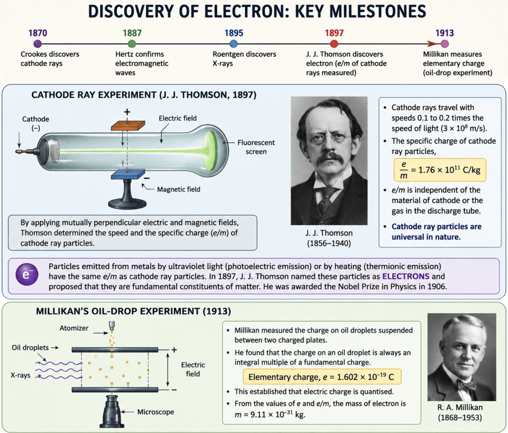
J. J. Thomson discovered the electron in 1897.
In a discharge tube containing gas at very low pressure, applying a high electric field caused a discharge and produced a fluorescent glow on the glass wall opposite the cathode.
William Crookes discovered cathode rays in 1870 and proposed that they were streams of fast-moving negatively charged particles.
J. J. Thomson later confirmed this idea experimentally using electric and magnetic fields and measured the speed and charge-to-mass ratio of these particles, which were later called electrons.
This passage continues the story of the discovery of the electron and explains important experimental results.
Main points
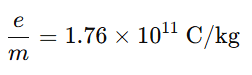
J. J. Thomson measured the specific charge of cathode ray particles:
The particles moved at speeds about .1C to .2C
where (c) is the speed of light.
The value of (e/m) was the same regardless of:
the metal used as the cathode,
or the gas inside the discharge tube.
This showed that cathode ray particles are universal and present in all matter.
Discovery of electrons from other experiments
Scientists also found that:
ultraviolet light falling on some metals caused emission of negative particles (photoelectric effect),
heated metals also emitted negative particles (thermionic emission).
The emitted particles had the same (e/m) ratio as cathode rays, proving they were identical.
Naming of the electron
In 1897, J. J. Thomson named these particles electrons and proposed that they are fundamental constituents of matter.
He received the Nobel Prize in Physics in 1906 for this work.
Millikan’s oil-drop experiment
In 1913, Robert Andrews Millikan measured the charge of the electron using the famous oil-drop experiment.
He found the elementary charge:
He also showed that electric charge is quantized, meaning charge always exists in whole-number multiples of this elementary charge.
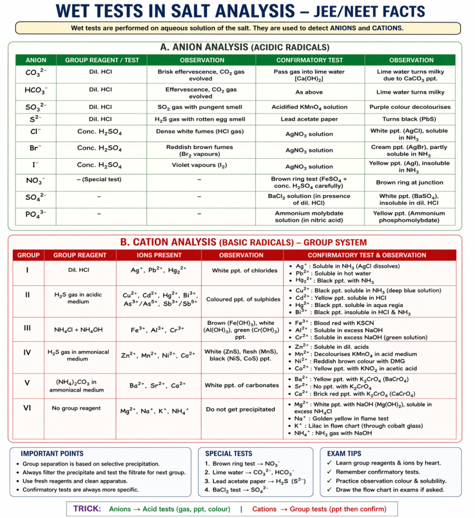
Salt analysis is a systematic method used to detect the cation and anion present in a given salt using dry and wet tests.
METHODS OF SALT ANALYSIS
1. Dry Tests
Flame test
Borax bead test
Charcoal cavity test
Cobalt nitrate test
Heating test
2. Wet Tests
Preliminary test (solubility test)
Anion analysis (acid radical tests)
Cation analysis (group analysis)
Confirmatory tests
3. Special Tests
Brown ring test
Lime water test
Chromyl chloride test
Lead acetate test
Nessler’s test
CATION GROUPS (Basic Radicals)
Group I (Dilute HCl group)
Ag⁺
Pb²⁺
Hg₂²⁺
Group II (H₂S in acidic medium)
Group II A (Copper group)
Cu²⁺
Cd²⁺
Hg²⁺
Pb²⁺
Bi³⁺
Group II B (Arsenic group)
As³⁺ / As⁵⁺
Sb³⁺ / Sb⁵⁺
Sn²⁺ / Sn⁴⁺
Group III (NH₄Cl + NH₄OH group)
Fe³⁺
Al³⁺
Cr³⁺
Group IV (H₂S in basic medium)
Zn²⁺
Mn²⁺
Ni²⁺
Co²⁺
Group V ((NH₄)₂CO₃ group)
Ba²⁺
Sr²⁺
Ca²⁺
Group VI (No group reagent)
Mg²⁺
Na⁺
K⁺
NH₄⁺
What is a Group Reagent?
A group reagent is a chemical reagent used in salt analysis to precipitate a specific group of cations together based on their similar properties.
Do anions have groups in salt analysis?
No, anions do not have a fixed “group system” like cations.
Why not?
Cations are grouped based on systematic precipitation using group reagents
Anions do not show such uniform precipitation behavior
Different anions require different specific tests
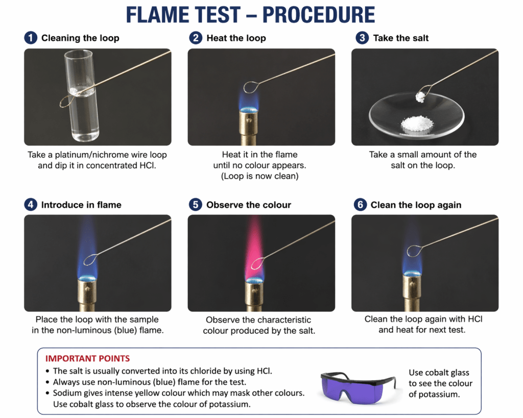
In qualitative analysis, dry tests are performed without dissolving the salt in solution. In a flame test, the salt (usually converted to its chloride) is directly heated in a flame, so it falls under dry tests. Flame test is done by heating a salt (on a clean wire loop with HCl) in a non-luminous flame and observing the characteristic color.
COMPLETE FLAME TEST LIST
1. Alkali Metals (Group 1) — Very important
Li⁺ (Lithium) → Crimson red
Na⁺ (Sodium) → Intense golden yellow
K⁺ (Potassium) → Lilac / pale violet (seen through cobalt glass)
Rb⁺ (Rubidium) → Red-violet
Cs⁺ (Caesium) → Blue-violet
2. Alkaline Earth Metals (Group 2)
Be²⁺ (Beryllium) → No colour
Mg²⁺ (Magnesium) → No colour
Ca²⁺ (Calcium) → Brick red (orange-red)
Sr²⁺ (Strontium) → Crimson red
Ba²⁺ (Barium) → Apple green
3. Transition & Post-Transition Metals
Cu⁺ / Cu²⁺ (Copper) → Bluish green
Pb²⁺ (Lead) → Bluish white (faint)
Tl⁺ (Thallium) → Bright green
Most others do NOT give a characteristic flame colour:
Fe²⁺ / Fe³⁺ → No colour
Co²⁺ → No colour
Ni²⁺ → No colour
Zn²⁺ → No colour
Mn²⁺ → No colour
Al³⁺ → No colour
4. Ions Showing NO Flame Colour (Important for elimination)
Be²⁺, Mg²⁺, Al³⁺
Zn²⁺, Fe²⁺/Fe³⁺, Co²⁺, Ni²⁺, Mn²⁺ Reason: emission not in visible region / weak excitation
IMPORTANT EXAM FACTS
1. Sodium Interference
Na⁺ gives very intense yellow
Masks all other colours
Solution: Use cobalt glass to detect K⁺
2. Why flame colour appears?
Due to electronic excitation and emission spectrum
3. Best observed for:
Volatile salts (usually chlorides)
What is a Wet Test?
A wet test is a method in salt analysis where the substance is first dissolved in a solvent (usually water or acid) and then tested using chemical reactions.
COLOURS OF CATION RADICALS
1. Coloured Ions (VERY IMPORTANT)
Cu²⁺ (Copper) → Blue solution
Fe²⁺ (Ferrous) → Pale green
Fe³⁺ (Ferric) → Yellow / brown
Ni²⁺ (Nickel) → Green
Co²⁺ (Cobalt) → Pink
Cr³⁺ (Chromium) → Green / violet
Mn²⁺ (Manganese) → Very pale pink
V³⁺ (Vanadium) → Green
Ti³⁺ (Titanium) → Purple
2. Colourless Ions (VERY COMMON MCQ)
Na⁺
K⁺
NH₄⁺
Mg²⁺
Ca²⁺
Ba²⁺
Sr²⁺
Al³⁺
Zn²⁺
3. Characteristic Precipitate Colours (IMPORTANT)
Ag⁺ → White ppt (AgCl)
Pb²⁺ → White ppt
Fe³⁺ → Reddish-brown ppt (Fe(OH)₃)
Fe²⁺ → Dirty green ppt (Fe(OH)₂)
Cu²⁺ → Blue ppt (Cu(OH)₂)
Ni²⁺ → Green ppt
Co²⁺ → Blue ppt (on heating)
Zn²⁺ → White ppt (Zn(OH)₂)
Al³⁺ → White gelatinous ppt
IMPORTANT EXAM POINTS
Colour is due to d–d transitions (transition metals)
d⁰ and d¹⁰ ions → colourless (Zn²⁺, Al³⁺ etc.)
Cu²⁺ always → blue solution (very important)
MOST ASKED
Blue solution → Cu²⁺
Green solution → Ni²⁺ / Fe²⁺ (trap!)
Pink → Co²⁺
Yellow/brown → Fe³⁺
SPECIAL REAGENTS – DETAILS
1. Nessler’s Reagent
Used for: Detection of NH₄⁺ (ammonium ion)
Composition
Alkaline solution of K₂HgI₄ (potassium mercuric iodide)
Observation
Brown colour / brown precipitate → NH₄⁺ present
Reaction idea
NH₃ (from NH₄⁺) reacts with Nessler’s reagent → coloured complex
One-line (viva)
Nessler’s reagent gives brown colour with ammonium ions.
2. FeSO₄ (Ferrous sulphate) – Brown Ring Test
Used for: Detection of NO₃⁻ (nitrate ion)
Reagents used
Fresh FeSO₄ solution
Concentrated H₂SO₄
Observation
Brown ring at junction of two layers
Key concept
Formation of nitrosyl complex
Important conditions
FeSO₄ must be fresh
H₂SO₄ added slowly along test tube wall
One-line (viva)
Brown ring test confirms presence of nitrate ion.
3. Copper Turnings Test
Used for: Detection of NO₃⁻ (nitrate ion)
Reagents used
Copper metal + conc. H₂SO₄
Observation
Brown fumes of NO₂ gas
Reaction idea
Nitrate gets reduced → NO₂ gas evolves
TEST FOR NICKEL (Ni²⁺) – KEY FACTS
1. Group Information
Group IV cation
Precipitated as NiS (black ppt) in basic medium (H₂S + NH₄OH)
2. Most Important Confirmatory Test
Dimethylglyoxime (DMG) Test
Reagent: Dimethylglyoxime (DMG) + NH₄OH
Observation:Bright red / rose-red precipitate
Compound formed: Nickel dimethylglyoxime complex
Very specific test for Ni²⁺
3. Reaction Idea
Ni²⁺ + DMG → Red complex (insoluble)
4. Other Supporting Facts
Ni²⁺ solution → Green colour
With NaOH → Green ppt (Ni(OH)₂)
With NH₄OH → forms complex (no ppt in excess)
What is Lake Test?
Lake test is used to detect Al³⁺ (aluminium ion) in salt analysis.
Principle
A lake is a coloured complex (dye + metal hydroxide) formed when a dye gets adsorbed on a gelatinous precipitate.
Procedure (short)
Add NH₄OH → forms Al(OH)₃ (white gelatinous ppt)
Add a dye (like alizarin / litmus)
Observation
Formation of a coloured “lake” (red/blue coloured ppt)
This confirms Al³⁺
Iodide of Millon’s base is involved in the Nessler’s reagent test.
In the test for NH4+ (ammonium ion), iodide of Millon’s base is involved in the Nessler’s reagent test.
Nessler’s Reagent (used for detection):
It contains alkaline solution of potassium tetraiodomercurate(II)
This is prepared using:
Mercury salts
Potassium iodide (KI)
KOH
During preparation, iodide of Millon’s base type species are formed.
Reaction with :
When ammonium ion is present:
Observation:
Brown precipitate forms This precipitate is called:
Iodide of Millon’s base
Charcoal Cavity Test (CCT)
The charcoal cavity test is a dry test in qualitative inorganic analysis used to detect certain metal ions (cations) by heating them in a small cavity made in charcoal.
Procedure:
A small cavity (hole) is made in a piece of charcoal
A pinch of the salt is placed in it
Heated strongly using a blowpipe flame
Sometimes mixed with sodium carbonate (Na₂CO₃)
Principle:
Charcoal acts as a reducing agent
On heating, metal compounds are reduced to metal or metal oxide
Different metals give characteristic residues / coatings
Observations (Important for exams):
Metal ion
Observation
Zn²⁺
Yellow when hot, white when cold (ZnO coating)
Pb²⁺
Yellow coating (PbO)
Cu²⁺
Red metallic bead (Cu)
Ag⁺
Shiny white metallic bead
Bi³⁺
Yellow coating
Borax Bead Test — Full Concept for JEE / NEET
This is one of the most important dry tests in qualitative analysis to identify transition metal ions.
What is Borax?
Borax = Sodium tetraborate
Principle (CORE CONCEPT)
On heating, borax decomposes to form boric anhydride which reacts with metal oxides:
Step 1: Heating borax
= boric anhydride (glassy mass)
Step 2: Reaction with metal oxide
Forms colored metaborate bead
Why color appears?
Due to transition metal ions
Caused by d–d electronic transitions
Flame Types (VERY IMPORTANT)
Flame type
Nature
Use
Oxidizing flame (O.F.)
Excess oxygen
Metal in higher oxidation state
Reducing flame (R.F.)
Less oxygen
Metal in lower oxidation state
Same metal → different color in OF & RF
Important Colors (JEE/NEET GOLD)
Metal ion
Oxidizing Flame
Reducing Flame
Cu²⁺
Blue-green
Red (Cu₂O / Cu)
Co²⁺
Deep blue
Deep blue
Cr³⁺
Green
Green
Fe³⁺
Yellow-brown
Bottle green
Mn²⁺
Amethyst (purple)
Colorless
Ni²⁺
Brown
Grey
V
Yellow
Green
Procedure (Short)
Take a platinum wire loop
Dip in borax → heat → forms colorless bead
Add salt → heat in O.F. & R.F.
Observe color
Key Points (Exam Focus)
Works mainly for transition metals
Bead = transparent glassy mass
Color depends on:
Metal ion
Oxidation state
Flame type
Cobalt always gives blue → very important MCQ
Common Mistakes Students Make
Ignoring reducing flame colors
Confusing Cu and Cr colors
Forgetting Mn becomes colorless in R.F.
Different Zones of Flame (very important for JEE/NEET)
1. Inner Zone (Dark Zone)
Location: Innermost part near burner
Color: Dark / almost invisible
Temperature: Lowest
Nature:Unburnt gases present (no combustion)
Contains mixture of fuel gas + air No practical use in tests
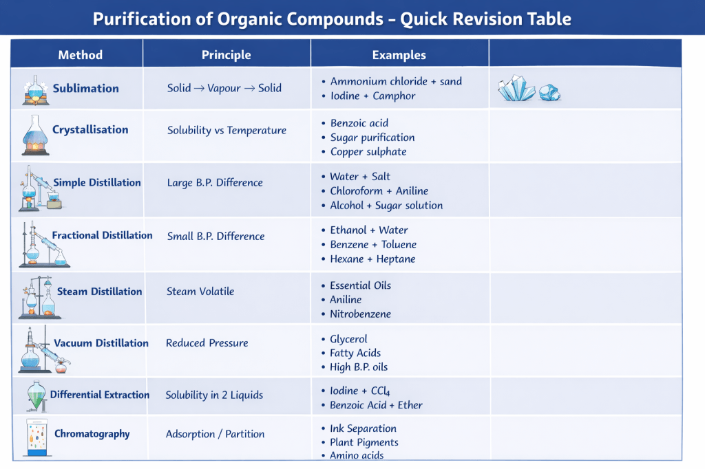
Extraction of essential oils From plants (e.g., clove oil, eucalyptus oil)
Example 2:
Aniline purification High b.p. but steam volatile
Example 3:
Nitrobenzene Separated using steam
Example 4:
Rose oil extraction (attar) Used in perfumes
Quick Comparison (Very Important)
Method
Condition
Example
Simple Distillation
Large b.p. difference
Water + salt
Fractional Distillation
Small b.p. difference
Ethanol + water
Steam Distillation
Heat-sensitive, steam volatile
Essential oils
Distillation Under Reduced Pressure (Vacuum Distillation)
Statement:
Distillation under reduced pressure → Glycerol + spent-lye
Why this method is used?
Some liquids:
Have very high boiling points
Decompose before boiling
Example: Glycerol
Principle
When pressure is reduced: ➡ Boiling point decreases
So, substance can distil at lower temperature without decomposition
Application in given case
Mixture:
Glycerol (high b.p., heat sensitive)
Spent-lye (impurities)
What happens?
Pressure ↓
Glycerol boils at lower temp
Vapours collected → pure glycerol
Impurities remain
Why not simple distillation?
❌ Glycerol decomposes at high temperature ✔ So vacuum distillation is required
🔹 More Examples
Example 1:
Glycerol purification (most common)
Example 2:
High boiling oils
Example 3:
Fatty acids
Example 4:
Petroleum residues
Quick Summary
Method
Used for
Simple distillation
Low b.p. liquids
Fractional distillation
Close b.p. liquids
Steam distillation
Heat-sensitive, steam volatile
Reduced pressure distillation
High b.p., decomposing liquids
Differential Extraction
When an organic compound is present in an aqueous medium, it is separated by shaking it with an organic solvent in which it is more soluble than in water. The organic solvent and aqueous solution should be immiscible so that they form two distinct layers which can be separated using a separating funnel. The organic solvent is later removed by distillation or evaporation to obtain the compound.
Concept: Differential (Liquid–Liquid) Extraction
Principle
Based on difference in solubility of a compound in two immiscible liquids
Usually:
Water (aqueous layer)
Organic solvent (like ether, benzene)
Key Conditions
✔ Two liquids must be immiscible ✔ Compound should be more soluble in organic solvent
Process (Separating Funnel)
Take mixture in separating funnel
Add organic solvent
Shake well
Allow layers to separate
Two layers form:
Upper layer (usually organic solvent)
Lower layer (water)
Separate layers
Evaporate solvent → get pure compound
Example
Example 1:
Iodine in water + carbon tetrachloride (CCl₄) → Iodine moves to organic layer
Example 2:
Benzoic acid from water using ether
Continuous Extraction
When used?
When compound is slightly soluble in solvent
Why?
Single extraction inefficient
Repeated extraction increases yield
Same solvent reused again and again
Advantages
Efficient separation
Better recovery of compound
Widely used in organic chemistry labs
Chromatography
What is Chromatography?
A separation technique where components of a mixture are separated based on their different affinities between two phases.
Principle
Based on distribution of components between two phases:
Stationary phase → fixed (solid or liquid)
Mobile phase → moving (liquid or gas)
Different substances move at different speeds → separation occurs
Types of Chromatography
1. Paper Chromatography
Stationary phase → paper
Mobile phase → solvent
Used for:
Ink separation
Plant pigments
2. Thin Layer Chromatography (TLC)
Stationary phase → silica gel layer
Faster and more accurate than paper chromatography
3. Column Chromatography
Column filled with adsorbent (silica/alumina)
Used for larger scale separation
4. Gas Chromatography (GC)
Mobile phase → gas
Used for volatile compounds
How Separation Happens?
Component with more attraction to stationary phase → moves slowly
Component with more attraction to mobile phase → moves faster
Example
Ink Separation
Black ink → separates into different colors on paper
Plant Pigments
Chlorophyll, carotene separated using chromatography
Important Term: Rf Value
Uses
✔ Separation of mixtures ✔ Purification ✔ Checking purity ✔ Drug testing ✔ Food analysis