
1. Gametogenesis
- Gametogenesis: The process of formation of gametes.
- Male gamete: Sperm.
- Female gamete: Ovum (female gamete).
- Primary sex organs: Testes in males and ovaries in females.
2. Spermatogenesis
Definition: The process of formation of spermatozoa (sperms) from spermatogonia in the testes.
- Begins at puberty.
- Occurs in the seminiferous tubules of the testes.
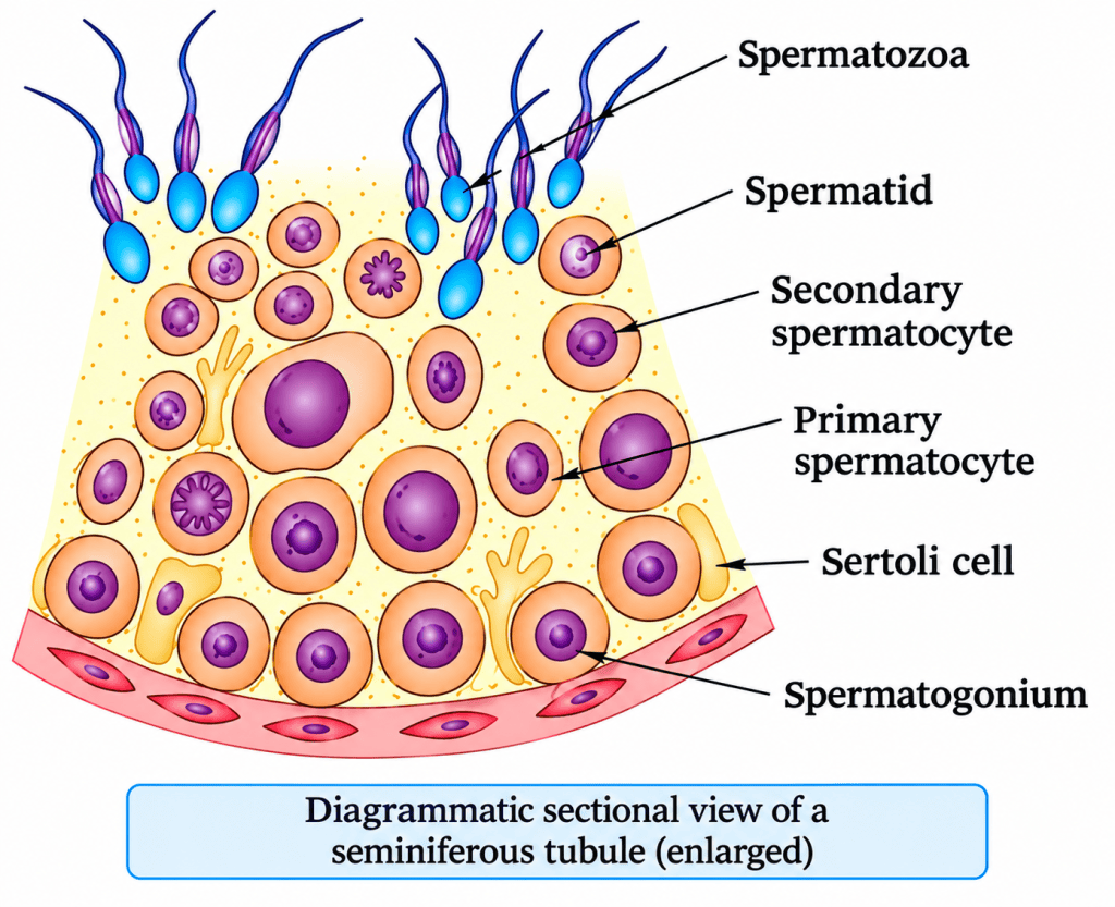
Steps of spermatogenesis
| Stage | Process | Chromosome number |
|---|---|---|
| Spermatogonium | Divides by mitosis | 46 (diploid) |
| Primary spermatocyte | Undergoes meiosis I | 46 (diploid) |
| Secondary spermatocytes (2) | Formed after meiosis I | 23 each (haploid) |
| Spermatids (4) | Formed after meiosis II | 23 each (haploid) |
| Spermatozoa (4) | Formed from spermatids | 23 each (haploid) |
Important terms
- Spermiogenesis: Transformation of spermatids into spermatozoa.
- Spermiation: Release of spermatozoa from Sertoli cells into the lumen of seminiferous tubules.
- Sertoli cells: Support developing sperm cells and provide factors that help spermiogenesis.
- Leydig cells: Produce and secrete androgens.
Hormonal control of spermatogenesis
| Hormone | Source | Function |
|---|---|---|
| GnRH | Hypothalamus | Stimulates the anterior pituitary to secrete LH and FSH |
| LH | Anterior pituitary | Acts on Leydig cells and stimulates androgen secretion |
| FSH | Anterior pituitary | Acts on Sertoli cells and stimulates secretion of factors that help spermiogenesis |
| Androgens | Leydig cells | Stimulate spermatogenesis |
Sequence to remember:
GnRH → LH and FSH
- LH → Leydig cells → Androgens → Spermatogenesis
- FSH → Sertoli cells → Factors supporting spermiogenesis
3. Structure of a Human Sperm
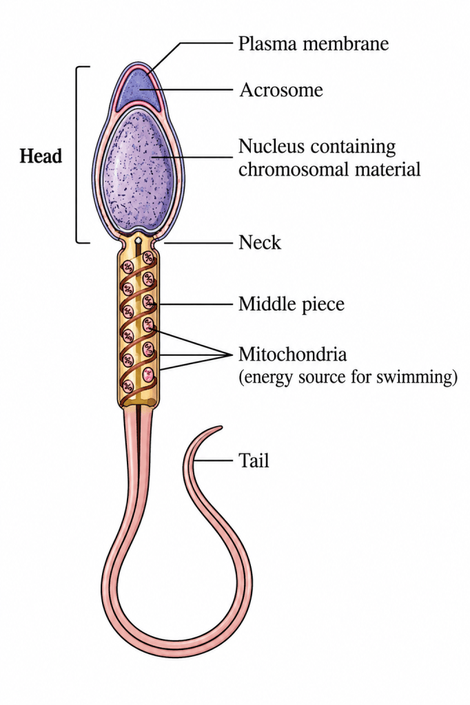
A sperm is a microscopic structure consisting of four parts.
| Part | Important features and functions |
|---|---|
| Head | Contains an elongated haploid nucleus |
| Acrosome | Cap-like structure covering the anterior portion of the head; contains enzymes that help fertilisation |
| Neck | Connects the head and middle piece |
| Middle piece | Contains numerous mitochondria that produce energy for movement |
| Tail | Helps in sperm motility |
| Plasma membrane | Encloses the entire sperm |
Important facts about human sperm
- Each sperm contains 23 chromosomes.
- The human male ejaculates approximately 200–300 million sperms during coitus.
- For normal fertility, according to the NCERT text:
- At least 60% of sperms must have normal shape and size.
- At least 40% must show vigorous motility.
4. Male Accessory Ducts and Glands
Accessory ducts
- Rete testis
- Vasa efferentia
- Epididymis
- Vas deferens
- Ejaculatory duct
- Urethra
Functions of accessory ducts:
- Transport sperms.
- Secretions of the epididymis, vas deferens, seminal vesicles and prostate are essential for sperm maturation and motility.
Accessory glands
- Seminal vesicles
- Prostate gland
- Bulbourethral glands
Important terms
- Semen: Sperms together with seminal plasma.
- Androgens: Maintain the functions of male accessory ducts and glands.
5. Oogenesis
Definition: The process of formation of a mature female gamete from oogonia.
Important facts
- Oogenesis begins during embryonic development.
- A couple of million oogonia are formed within each fetal ovary.
- No new oogonia are formed and added after birth, according to NCERT.
- Primary oocytes remain temporarily arrested in prophase I of meiosis.
- At puberty, approximately 60,000–80,000 primary follicles remain in each ovary.
Stages of oogenesis and follicular development
| Stage | Description |
|---|---|
| Oogonium | Diploid female germ cell |
| Primary oocyte | Enters meiosis I during embryonic development and gets arrested in prophase I |
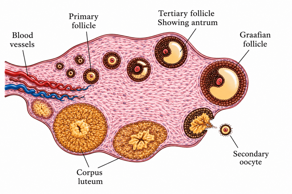
| Primary follicle | Primary oocyte surrounded by a layer of granulosa cells |
| Secondary follicle | Primary follicle develops more layers of granulosa cells and a new theca |
| Tertiary follicle | Contains a fluid-filled cavity called the antrum |
| First meiotic division | Produces a large haploid secondary oocyte and a tiny first polar body |
| Graafian follicle | Mature follicle containing the secondary oocyte |
| Ovulation | Release of the secondary oocyte from the ovary |
Important structures of the ovarian follicle
- Granulosa cells: Surround and support the developing oocyte.
- Theca interna: Inner layer of the theca.
- Theca externa: Outer layer of the theca.
- Antrum: Fluid-filled cavity of the tertiary follicle.
- Zona pellucida: Membrane that surrounds the secondary oocyte.
Meiotic division during oogenesis
- The first meiotic division is unequal.
- It produces:
- One large haploid secondary oocyte.
- One tiny first polar body.
- The secondary oocyte retains most of the nutrient-rich cytoplasm.
- The first polar body may divide further or degenerate; NCERT states that this is not certain.
- The Graafian follicle ruptures and releases the secondary oocyte during ovulation.
Why does the secondary oocyte retain most of the cytoplasm?
It ensures that the female gamete retains sufficient cytoplasm, organelles and stored nutrients to support early development after fertilisation.
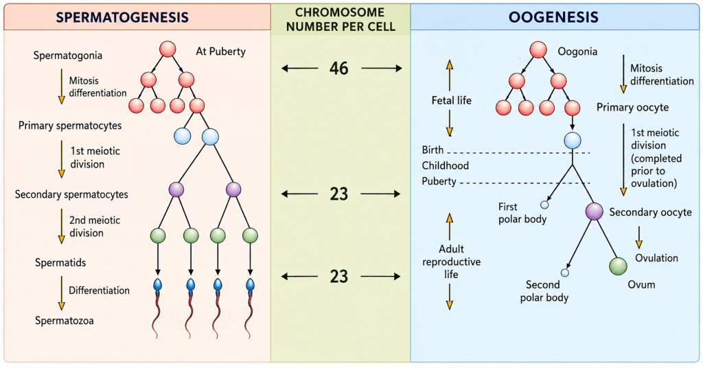
6. Differences Between Spermatogenesis and Oogenesis
| Feature | Spermatogenesis | Oogenesis |
|---|---|---|
| Site | Seminiferous tubules of testes | Ovaries |
| Begins | At puberty | During embryonic development |
| Germ cells | Spermatogonia | Oogonia |
| Growth phase | Comparatively short | Long and pronounced |
| Cytoplasmic division | Equal | Unequal |
| Products of meiosis | Four haploid spermatids | One large secondary oocyte and polar body after meiosis I |
| Functional gametes | Four sperms from one primary spermatocyte | One functional female gamete from one primary oocyte |
| Nature | Continuous after puberty | Cyclic release of secondary oocytes during reproductive life |
| Gamete size | Small | Large |
| Motility | Sperm is motile | Female gamete is non-motile |
7. Quick Revision: Must-Remember Facts
46 → 23 chromosomes
Meiosis I reduces the chromosome number from diploid to haploid.
Spermiogenesis vs spermiation
Spermiogenesis = spermatids transform into spermatozoa.
Spermiation = spermatozoa are released into the lumen of seminiferous tubules.
LH vs FSH
LH acts on Leydig cells; FSH acts on Sertoli cells.
Oogenesis begins before birth
Primary oocytes are arrested in prophase I.
Antrum and zona pellucida
Antrum = fluid-filled cavity of the tertiary follicle.
Zona pellucida = membrane surrounding the secondary oocyte.
Ovulation
Release of the secondary oocyte from the Graafian follicle.

